
Some of my science-based artwork and artistic representations of scientific results.
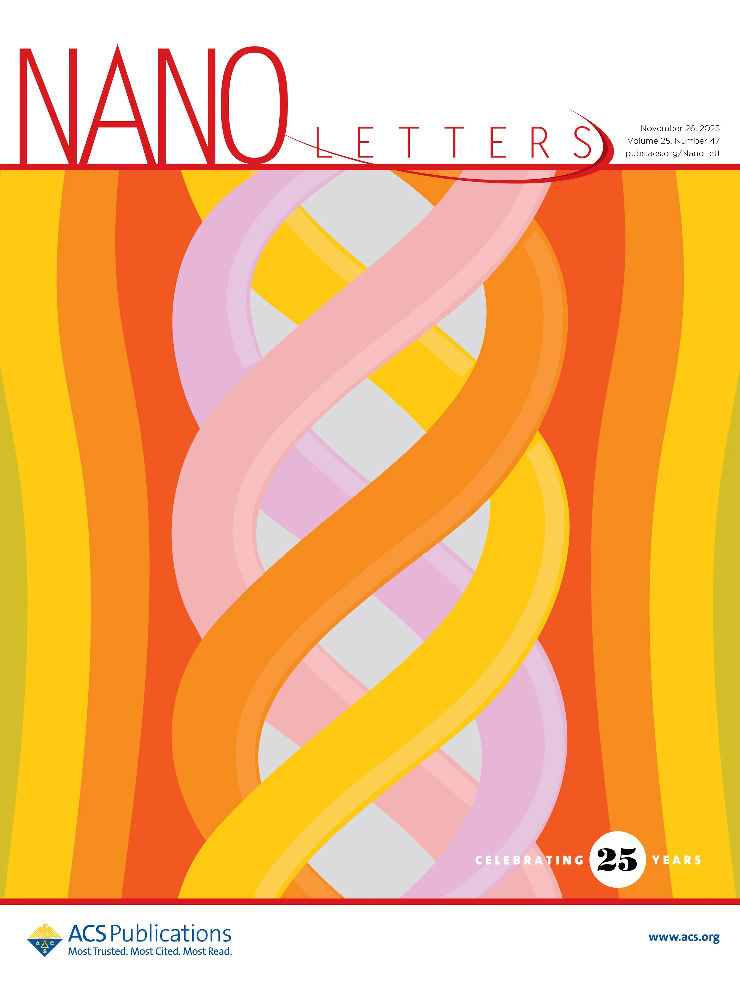
Paranemic crossover DNA. Multi-stranded DNA structures that are connected by strand crossovers can reassociate at specific temperatures to form double helical structures. One model structure, the paranemic crossover (PX) DNA is a structure that contains four DNA strands (shown in different colors in the center) organized in two double helical domains. Cover art designed and created by Arun Richard Chandrasekaran.
DNA rotary device. The image shows the two double helical domains of a PX-JX device (looking through helix axis), and crossovers between the two domains. A combination of RNase and DNA turns the bottom domains by 180 degrees. Cover art designed and created by Arun Richard Chandrasekaran.
Fluorescent intercalators. An image of a “kolam”, a traditional art from the state of Tamil Nadu in India. A kolam is typically composed of a series of dots and lines. Here, the lines represent DNA strands folded into a nanostructure and the dots represent intercalating molecules bound to the DNA nanostructures. In this work, fluorescent intercalators such as ethidium bromide are used to analyze the biostability of DNA nanostructures. Cover art designed and created by Arun Richard Chandrasekaran.
Toehold clipping. DNA nanostructures can be reconfigured by toehold-based DNA strand displacement. By incorporating RNA toeholds (blue) or toeholds with photocleavable linkers (orange) into DNA tetrahedra, strand displacement can be controlled using a process called “toehold clipping”, using a ribonuclease (yellow) or ultraviolet light (violet) as stimuli, respectively. Cover art designed and created by Arun Richard Chandrasekaran.
Encoding information in DNA nanoswitches. DNA nanoswitches with tunable loop sizes (bottom panel) encode information as 5-bit binary codes using nucleic acid inputs (middle panel). Information is decrypted using enzymes and read out using gel electrophoresis (top panel). Cover art designed and created by Arun Richard Chandrasekaran.
Fluorescent aptaswitch for detection of lead ions. A newly designed DNA aptamer switch (keyhole on door) can reconfigure when binding to lead ions (purple spheres). The aptamer switch (aptaswitch) is used to detect lead ions using the fluorescent signal obtained on reconfiguration (open aptaswitches on other side of door). Cover art designed and created by Arun Richard Chandrasekaran.
Aptamers for viral detection and inhibition. Viral infections are a major cause of disease in humans, plants, and animals. Aptamers (shown in an assembly line on a conveyor belt) are being developed as molecular tools for creating diagnostics and therapeutics for viral infections. Cover art designed and created by Arun Richard Chandrasekaran.
RNA purification using DNA nanoswitches. DNA nanoswitch technology enables benchtop purification of specific RNA sequences from biological extracts with high recovery and purity. The technique is competitive with beads-based methods and can be paired with LC/MS for identifying RNA modifications in specific RNAs. The cover image shows a girl fishing using a DNA nanoswitch “hook” that binds to an RNA sequence (shown as a single-stranded RNA folded into a fish), used to purify specific sequences of RNAs from biological samples. Cover art designed and created by Arun Richard Chandrasekaran.
Nuclease resistance of DNA nanostructures. Nanostructures built from DNA are being applied in biosensing, cell modulation, bioimaging and drug delivery. But the sensitivity of these structures to nucleases present in the physiological environment is an impediment. Strategies that increase the nuclease resistance of DNA nanostructures while retaining their functions are thus of great interest as are methods to evaluate resistance and quantify stability. Image: Arun Richard Chandrasekaran & Carl Conway; Design: Carl Conway.
Detecting Alzheimer’s microRNAs. MicroRNAs (shown as colored single strands in the brain) have emerged as likely disease regulators and biomarkers for Alzheimer's disease, now implicated as having roles in several biological processes related to progression of the disease. In this work, we use programmable DNA nanoswitches (shown as a double stranded DNA with “capturing” strands) for low-cost, non-enzymatic, single-step detection of Alzheimer’s associated microRNAs. Cover art created for ACS Sensors by Arun Richard Chandrasekaran.
Parallel control. A scientist walking on a DNA nanoswitch rope juggling three triggers (nucleic acid, light, ribonuclease) that can orthogonally reconfigure the nanoswitch into different conformational states. Cover image created for the journal Biochemistry by Arun Richard Chandrasekaran.
Count your syns. Characterization of DNA G-quadruplex topologies with NMR chemical shifts. Cover image created for Journal of Physical Chemistry Letters by Arun Richard Chandrasekaran.
Nuclease degradation analysis using gel electrophoresis. This protocol describes the use of gel electrophoresis to characterize nuclease resistance of DNA nanostructures using paranemic crossover (PX) DNA motif as an example (shown as a model in bottom right). By analyzing nuclease digestion (image hanging on left wall) of different nanostructures, one can obtain relative biostability enhancement factors (BioEF). Cover art designed and created by Arun Richard Chandrasekaran.
DNA-based graphical displays. DNA-based devices are useful in biosensing, molecular computation, and directed chemical reactions. These molecular processes can be readout using programmable graphical displays on nanoscale (DNA origami) or macroscopic (multi-well plates) platforms. Cover art designed and created by Arun Richard Chandrasekaran.
Ribonuclease-responsive DNA nanoswitches. Pac-Man analogy of ribonuclease triggered DNA nanoswitches: The ribonuclease (yellow Pac-Man) can act on DNA nanoswitches locked by RNA strands (shown as orange "Ghost" in the center) causing the switches to open. This RNase responsive process is used in in detecting RNases, screening RNase inhibitors and as molecular erasers in information encoding using DNA. Cover image created for Cell Reports Physical Sciences by Arun Richard Chandrasekaran.
How to perform miRacles. In the famous tortoise and the hare fable, we are taught that “slow and steady” wins the race. A DNA nanoswitch detection approach (“miRacles” assay) uses the slow and steady migration of a looped DNA as a detection signal to identify the presence of specific microRNAs (top band in right gel lane). This DNA nanotechnology approach provides low‐cost, non‐enzymatic, and direct detection of microRNAs by causing a conformational change in a DNA nanoswitch. Cover art designed and created by Arun Richard Chandrasekaran.
A superhero suit for DNA nanostructures. Applications of DNA nanostructures in diagnostics, therapeutics and biomolecular analysis. Cover art designed and created by Arun Richard Chandrasekaran.
Faux product packaging of DNA nanotechnology toolkit for microRNA detection. Technologies based on nanostructures such as DNA tetrahedra, DNA origami and DNA devices may offer “new and improved” features when compared with “leading brands” q-PCR, microarray and Northern blotting. Cover art designed and created by Arun Richard Chandrasekaran.
TERS for DNA detection. Tip-enhanced Raman imaging of single-stranded DNA with single base resolution. Cover image created for Journal of the American Chemical Society (JACS) by Arun Richard Chandrasekaran.
DNA based memory device. A stylistic portrayal of the DNA nanoswitch used to demonstrate a rewriteable 5-bit memory system. Looped conformations of DNA nanoswitches (center piece) collectively provide 1's and unlooped conformations are 0's, encoding the words "Hello World", with a gel electrophoresis readout. Cover art designed and created by Arun Richard Chandrasekaran.
Paranemic crossover (PX) DNA. A motif in DNA nanotechnology used to connect topologically closed molecules, to assemble a 3D object, and to create 2D DNA crystals. Cover art designed for the journal Chemical Reviews by Arun Richard Chandrasekaran.
A third strand for DNA nanotechnology. Triplex-forming oligonucleotides are starting to find room in DNA nanotechnology and offer the ability to create self-assembled lattices, reconfigurable objects and nanoscale machines and devices. Cover image created for Nucleic Acids Research by Arun Richard Chandrasekaran.
Sticky ends. DNA structures can be assembled using complementary single stranded regions called sticky ends. Image created by Arun Richard Chandrasekaran.
DNA microcrystals. DNA nanostructures can assemble into designer DNA crystals. Image created by Arun Richard Chandrasekaran.